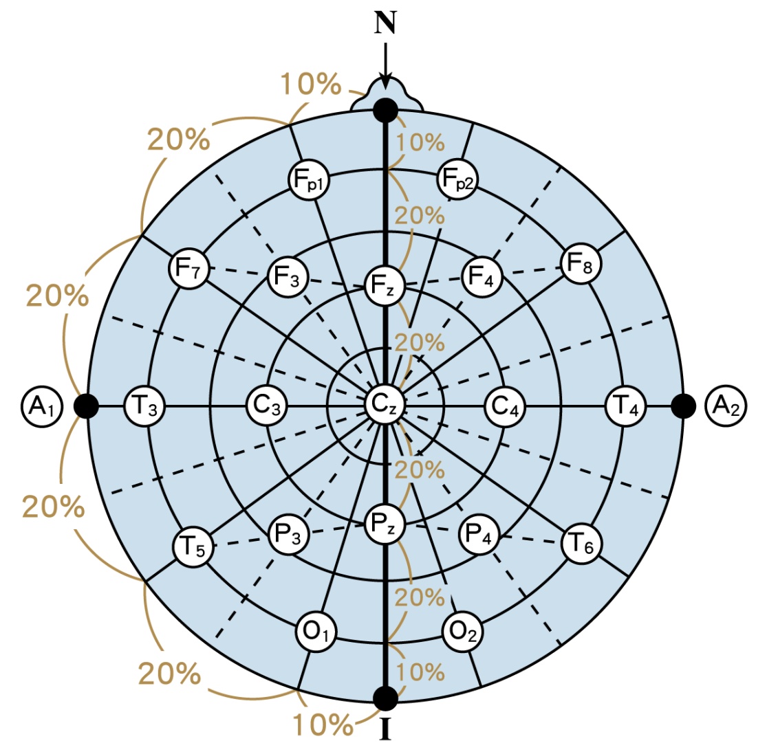
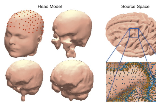
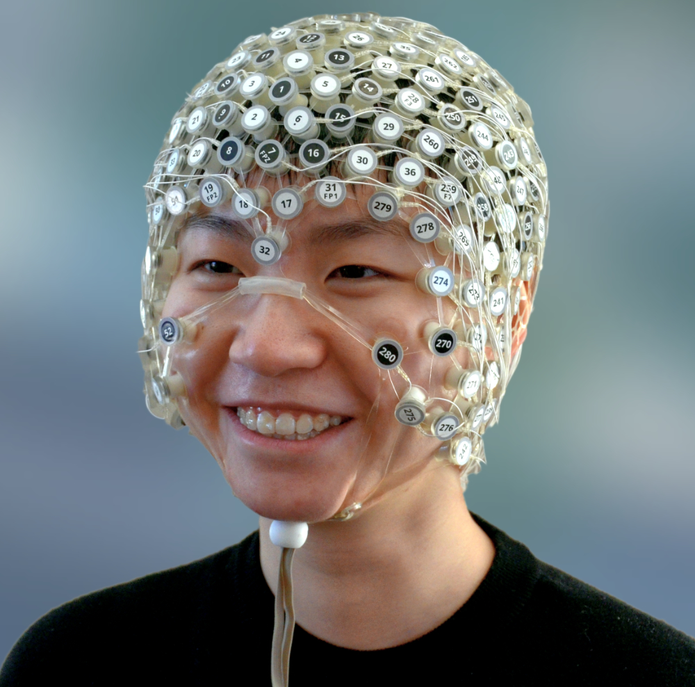
dEEG electrode distribution across the scalp is dense compared to the standard International 10-20 system used in many research and clinical settings (see below).
Glossary
- Electroencephalography (EEG)
Developed in 1929, EEG electrodes are placed on the human scalp to record electric signals from the top layers of the brain. The International 10-20 system established standard electrode locations across the scalp. Note the position of electrodes F3 and F4 on either side of the head.
- EEG Frequencies
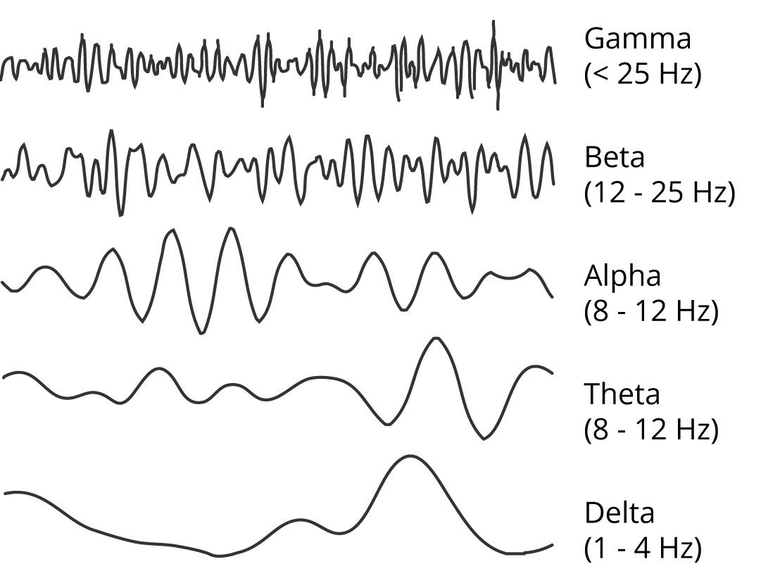
The brainwaves collected from the human scalp typically appear in five canonical frequencies which are correlated with resting eyes open or eyes closed, and during various tasks (movement or thinking).
- Electromyography (EMG)
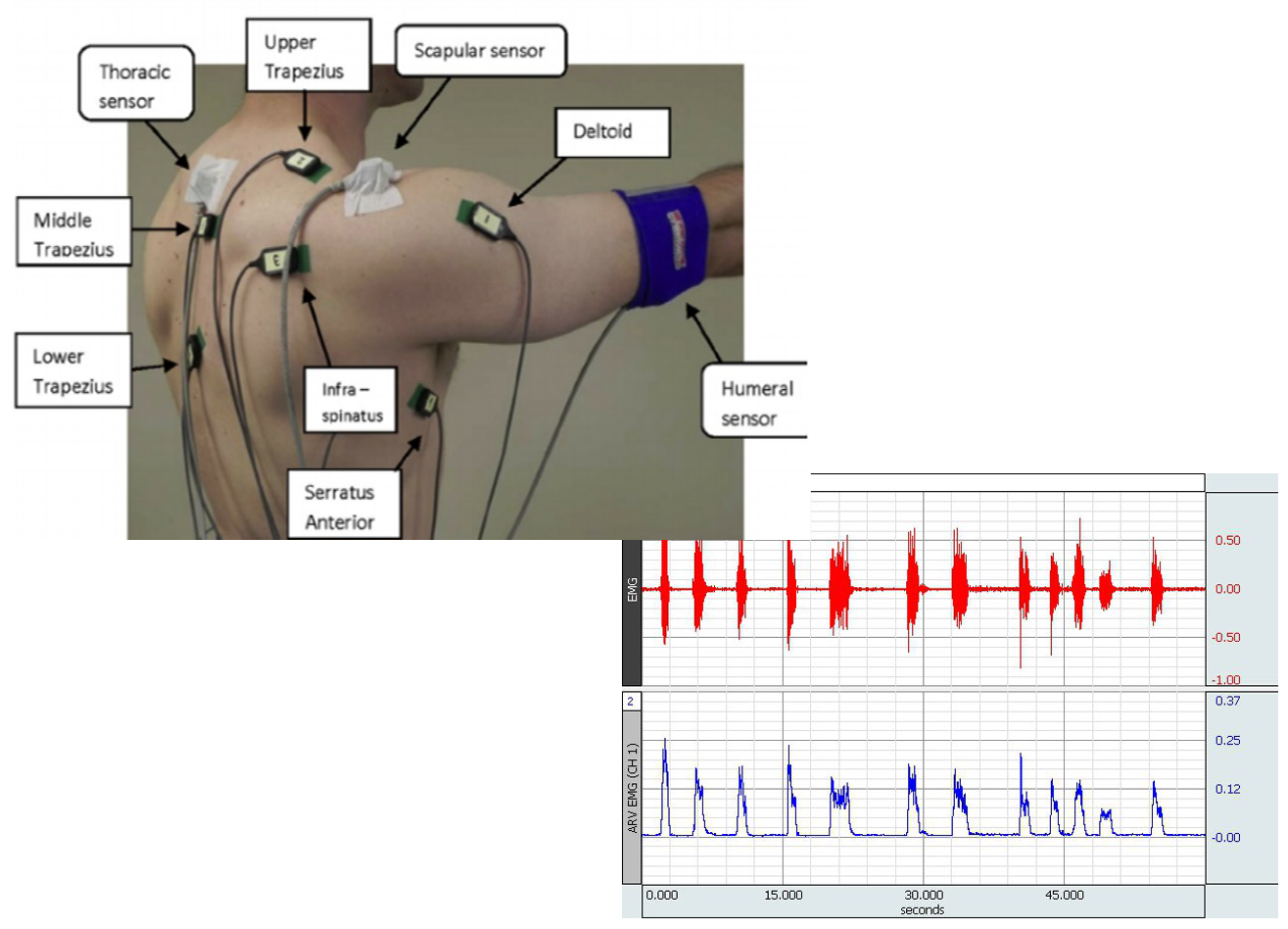
EMG measures electrical activity in selected surface muscle groups. EMG signals provide information on amount and frequency of muscle activity.
- Geodesic Photogrammetry System (GPS)
This system utilizes 11 video cameras to record the positions of each electrode.
- Head modeling
The human head is composed of a number of different tissues, each with its own electrical conductivity properties including brain tissue, connective tissue (e.g., dura mater) and bone (the skull). In addition, each person’s brain has a generally similar shape compared to other people, however, each person’s brain does have unique variations on the general shape. EEG head modeling techniques allow processing of EEG signals that take tissue conductivity properties into account, as well as positioning of electrodes for recording or weak electrical current injection that optimizes data collection accuracy and intervention effects.
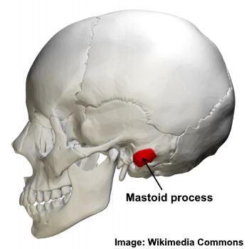
- Mastoid Location
The mastoid bones are located on both sides of the head just behind and below the ear.
- Memory
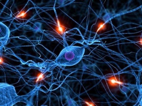
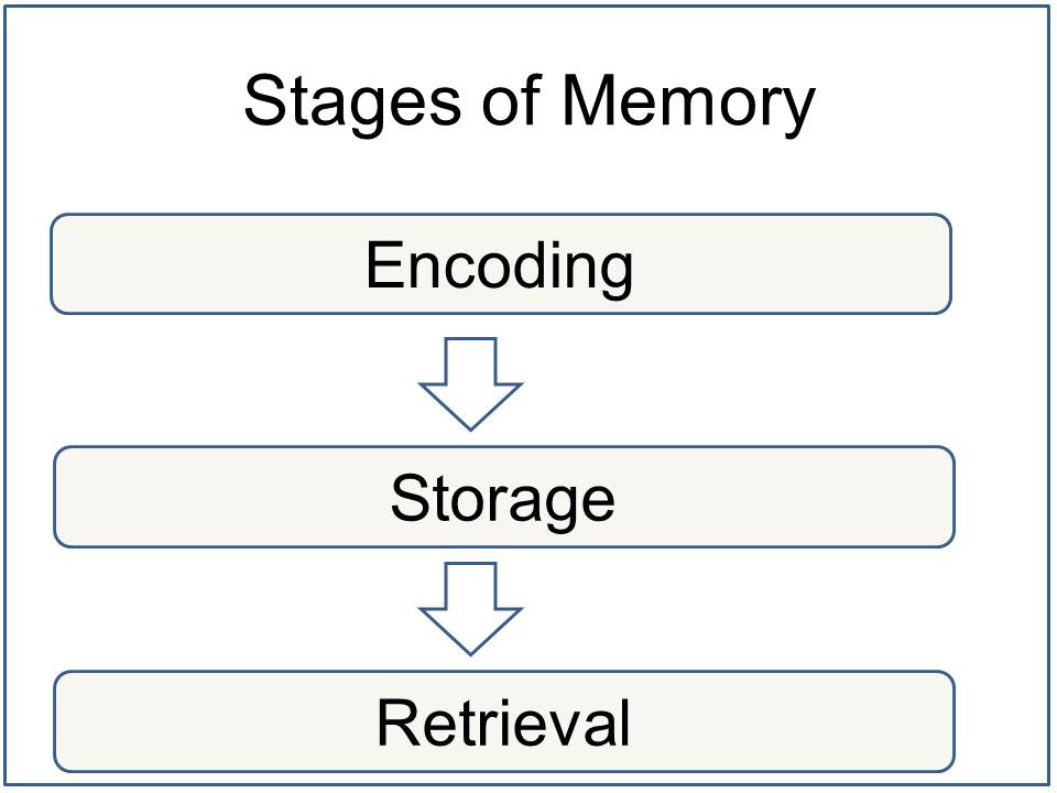
Memory begins between neurons at places called synapses which are structures in the neuron membrane that receive information from other neuron synapses. Synapses are modified to permit easy sharing of information or ‘decommissioned’ such that transmission is lessened or stopped at that synapse. Memory is created in stages, as seen above. Different kinds of memory are associated with different brain regions.
- Declarative Memory
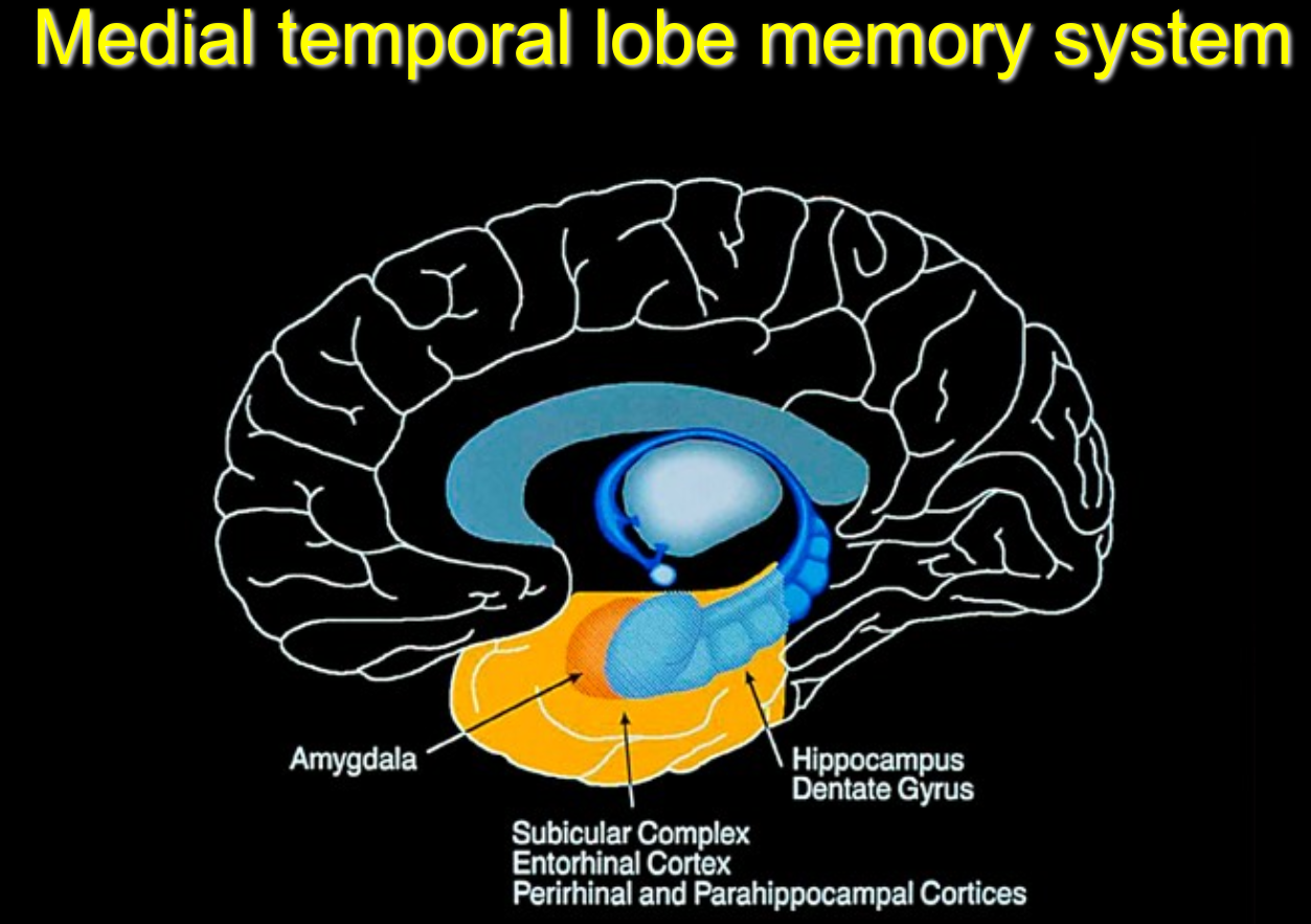
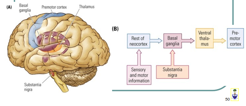
Declarative (explicit) memories are those that can be reported verbally. This includes autobiographical and factual information. This type of memory is associated with activity in deep areas of the brain in what has been called the limbic system (medial temporal lobe).
- Procedural Memory
Procedural (implicit) memory encodes and permits motor behaviors that do not require conscious awareness to perform such as walking, running, and riding a bike.
- Memory consolidation
Memory consolidation (storage) is thought to occur during sleep when synapses are either strengthened or decommissioned.
- Sleep
Sleep is a state common to all mammals. It is characterized by loss of normal consciousness and dreaming. It is required for life and health. Memories are thought to be consolidated during sleep.
- Sleep Stages
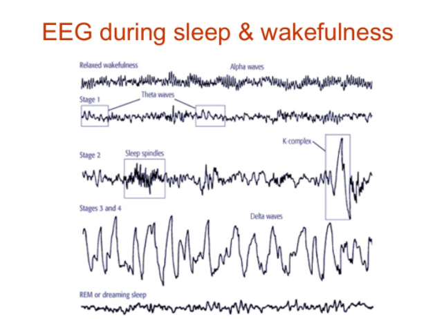
Sleep in humans proceeds through standard stages. These stages can be visually identified in EEG data traces. Slow wave sleep is termed non-rapid eye movement (NREM) sleep stages 3-4. It is thought memories are consolidated during NREM slow wave sleep. Rapid eye movement (REM) sleep can occur throughout the night and has been associated with dreaming.
- Source Localization
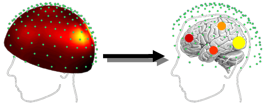
Use of mathematical signal analysis to determine the brain sources of electrical activity recorded at the scalp.
- Transcranial Direct Current Stimulation (tDCS) Electrodes
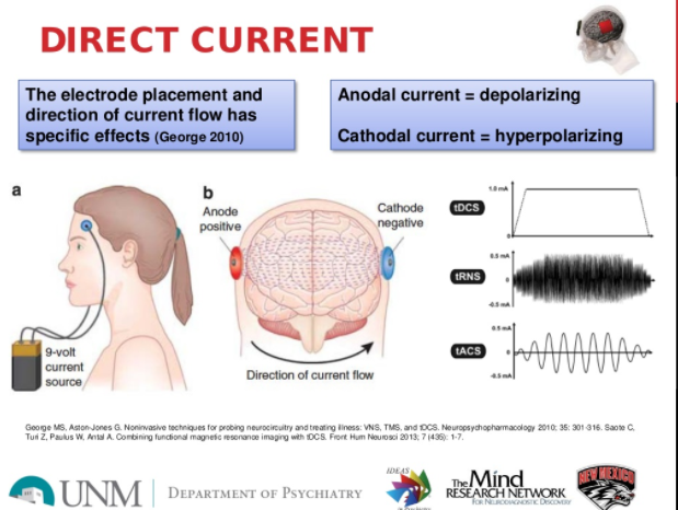
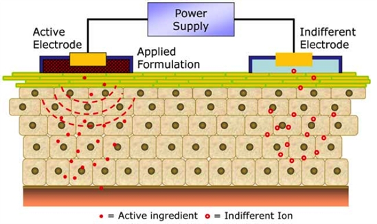
In typical tDCS neurodulation there are two electrodes, anodal (positive) and cathodal (negative). Anodal stimulation has been associated with increasing neuron probability of signaling and cathodal stimulation has been associated with lessening neuron probability of signaling.
