Increasingly, computation is extending mathematics as the language of science.
The BEL training integrates the interdisciplinary framework of neuroinformatics with the basic principles of human brain electrophysiology and neuroimaging.
The Brain Electrophysiology Laboratory mixes industry and academic cultures to weave together science and technology training.
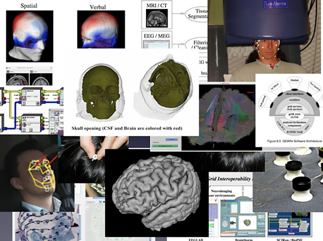
This weaving of science and technology requires cross training among students, scientists, and engineers in multiple basic disciplines, including psychology, neurophysiology, neuroanatomy, computational science, and several physical and chemical technologies. The collaborative team at BEL has come together to work on some of the complex issues facing the neuroimaging field.
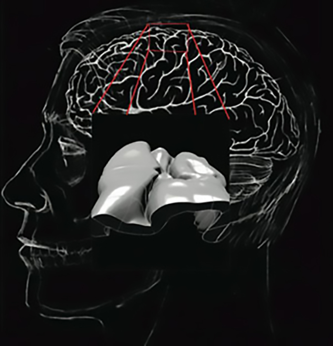
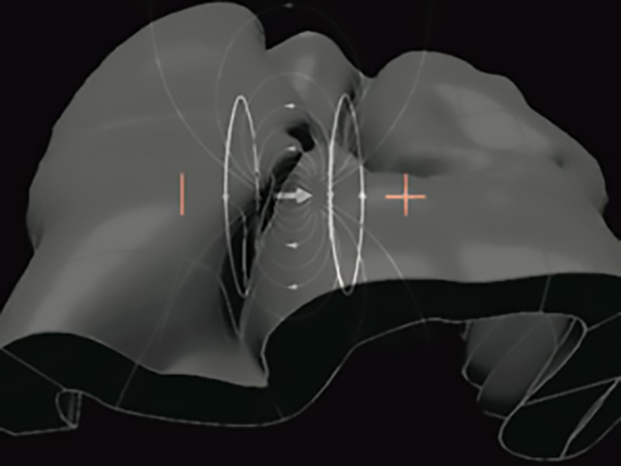
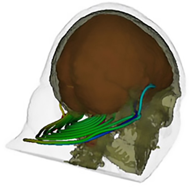
The perspectives from multiple disciplines are then applied in multiple areas, including medicine, education, and human performance. One area this wealth of knowlege has helped to shed light on is the electrical fields of the brain.
The principles of dense array EEG involve physical and physiological concepts. In the laboratory, BEL scientists, graduate students, research assistants, and postdocs work on a regular basis with BEL engineers and computer scientists from the NIC to share perspectives and techniques between disciplines.
Increasingly, computation is extending mathematics as the language of science.
The BEL training integrates the interdisciplinary framework of neuroinformatics with the basic principles of human brain electrophysiology and neuroimaging.
Ferree, T. & Tucker, D. M. (1999). Development of high-resolution EEG devices. International Journal of Bioelectromagnetism, 1, 4-10.
Ferree, T. C., Eriksen, K. J., & Tucker, D. M. (2000). Regional head tissue conductivity estimation for improved EEG analysis. IEEE Trans Biomed Eng, 47(12), 1584-1592.
Luu, P. & Tucker, D. M. (2003). Self-Regulation and the Executive Functions: Electrophysiological Clues. In A. Zani & A. M. Preverbio (Eds.), The cognitive electrophysiology of mind and brain. (pp. 199-223). San Diego: Academic Press.
Tucker, D. M. (1993). Spatial sampling of head electrical fields: the geodesic sensor net. Electroencephalogr Clin Neurophysiol, 87(3), 154-163.
Tucker, D. M., Liotti, M., Potts, G. F., Russell, G. S., & Posner, M. I. (1994). Spatiotemporal analysis of brain electrical fields. Human Brain Mapping, 1, 134-152.